


This article was automatically translated from the original Turkish version.
+1 More
Aphantasia is defined as the inability of individuals to form visual imagery in their minds. This term was first widely introduced in 2015 by science researcher Adam Zeman, and since then it has been studied as a neurological phenomenon. Individuals with aphantasia cannot generate any visual images when they close their eyes or attempt to think about a mental image. This condition does not only affect visual imagination but may also influence memory, creative thinking, and problem solving processes in distinct ways.
Aphantasia is a neurological condition that limits individuals’ ability to work with mental imagery. It arises from differences in how the brain processes and stores visual information. People with aphantasia can typically perceive the external world but are unable to form mental pictures or use visual imagination. Some research has revealed that this phenomenon was previously unrecognized and was mostly identified through personal accounts. Therefore, understanding and defining aphantasia has gained greater clarity only in recent years through neuroscientific studies.
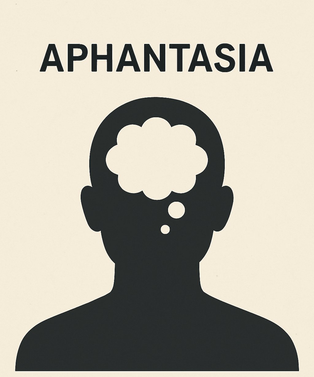
A visual representation of aphantasia (generated by Artificial Intelligence.)
Aphantasia was first mentioned in scientific literature in the 1880s by Francis Galton during his research on visual imagination. Galton conducted a series of surveys to test the vividness of individuals’ mental imagery. Participants were asked to mentally picture a landscape or an object, and most reported being able to clearly visualize these images. However, some participants stated they could not form any images at all or could only perceive very faint mental pictures. Galton noted this phenomenon and formed the first impressions that some individuals lacked the capacity to generate mental imagery. Yet Galton did not fully define the condition or label it as a distinct phenomenon.
The term “aphantasia” was scientifically defined in 2015 through a study conducted by Adam Zeman and his colleagues. While working as a neurologist at the University of Exeter, Zeman made significant findings regarding mental imagery. During this period, the term aphantasia began to be used to mean “mind blindness” or “lack of visual imagination.” Zeman’s research demonstrated that the inability to form mental images was not limited to visual imagery alone but could also involve the absence of other sensory imagery. This study was the first major research to define aphantasia as a scientific concept.
Following its definition, further research revealed that the number of individuals experiencing aphantasia is higher than previously thought. In their 2015 publication titled “Visual Imagery Deficiency,” Zeman and his team provided the first scientific definition of aphantasia and applied the first standardized tests. The study showed that aphantasia is not a rare condition but may be a widespread phenomenon affecting many people around the world.
These studies also began to provide important clues about the neurological basis of aphantasia. Individuals with aphantasia showed reduced activity in brain regions responsible for visual processing compared to those with typical visual imagination. These findings suggest that visual imagination is governed by specific brain areas and that aphantasia may stem from functional differences in these regions.
Aphantasia is a neurological condition that demonstrates the significant impact of brain structures and functions on visual imagination. Several brain regions play roles in generating and processing visual imagery. Aphantasia is associated with functional differences or deficits in these regions. Neuroscientific studies on these areas have helped us understand the neurological foundations of aphantasia.
Visual imagination arises through the cooperation of several key brain regions. Aphantasia is thought to be linked to functional differences or disconnections in these areas. The primary regions responsible for generating visual imagery are the occipital lobe and the parietal lobe. These regions play a critical role not only in visual perception, processing, and memory but also in the creation of mental images.
In individuals with aphantasia, these brain regions show lower levels of activity when compared to those with typical visual imagination. This may hinder the brain’s ability to process visual imagery. Additionally, differences are believed to exist in the functional connections involved in encoding and recalling visual images. Furthermore, aphantasia may also involve altered functioning in brain regions related to memory, such as the hippocampus. The hippocampus is a structure involved in processing visual memory and perception and in storing memories. A deficiency in its capacity to process visual imagery is one of the core features of aphantasia.
Visual imagination is directly linked to the brain’s ability to process visual information. Neurologically, these processes are typically managed by regions such as the occipital lobe and the temporal lobe. In aphantasia, the activity of brain regions responsible for visual imagination is weakened or absent. At the same time, individuals with aphantasia can perceive visual information from the external world but lack the ability to process it into mental images.
Visual imagination also involves planning and memory-related functions directed by the prefrontal cortex. This brain region plays a crucial role in supporting creative processes of imagination and processing stored images. Individuals with aphantasia may also exhibit weakened functional connections in these areas. Moreover, it has been suggested that aphantasia is not merely a lack of visual imagination but may also affect cognitive processes. In particular, difficulties may arise in tasks involving problem solving, creative thinking, and mentally reconstructing memories. There may also be disruptions in the functional connections that organize and guide these processes and manage visual information.
Aphantasia is a condition characterized by deficiencies in generating mental imagery, and its diagnosis typically relies on self-reports of personal experiences and various psychological tests. The diagnostic process for aphantasia includes a series of steps involving individual awareness of visual imagination deficits and neurological assessments. Accurate diagnosis enables a better understanding of aphantasia’s specific symptoms and more precise evaluation of personal experiences.
The diagnosis of aphantasia is primarily based on clinical evaluation and self-reporting. During this process, experts assess how individuals experience their imagination and mental imagery. The absence of visual imagination is usually defined by the individual’s own account and is generally accepted as an independent phenomenon, not necessarily linked to other psychological or neurological conditions. However, the diagnostic process requires consideration of other neurological and psychological conditions to better understand the effects of aphantasia.
The most common method for diagnosing aphantasia is to question and test individuals’ ability to form mental images. These tests aim to determine how imagination functions, where difficulties occur, and to what degree mental images can be formed. Such assessments are essential for understanding the scope of aphantasia.
The diagnostic process for aphantasia is not limited to tests evaluating the absence of visual imagination. Diagnosis also involves reviewing the individual’s psychological and neurological status. To gain further insight into the neurological basis of aphantasia, techniques such as functional brain imaging may be employed.
Neuroimaging Methods: Techniques such as fMRI (functional magnetic resonance imaging) are used to examine the neurological processes related to brain structures in aphantasia. These methods measure activity in brain regions and allow researchers to observe how areas involved in visual imagination function differently in individuals with aphantasia.
Psychological Assessment: An individual’s overall cognitive functioning is also considered. Psychological evaluations assess memory, attention, sensory perception, and other cognitive functions. Such tests are important to distinguish aphantasia from other mental disorders, as similar symptoms may be associated with certain neurological conditions.
Clinical Interviews and Questionnaires: The diagnostic process for aphantasia is supported by clinical interviews and questionnaires that provide a deep understanding of individuals’ experiences. Experts use targeted questions to identify specific challenges individuals face with visual imagination, thereby revealing the unique characteristics of aphantasia in each case.
Various tests and measurement methods are used in the diagnostic process of aphantasia to evaluate visual imagination. These tests aim to determine individuals’ capacity to form mental images. The most commonly used methods include:
Vividness of Visual Imagery Questionnaire (VVIQ): The VVIQ is a frequently used questionnaire for diagnosing aphantasia. This test asks individuals to imagine specific objects or scenes and then rate the vividness and clarity of the mental images they form. Participants are given a scale consisting of a series of questions to evaluate the vividness of their mental imagery. Results indicate the intensity and clarity of visual imagery. Individuals with aphantasia typically score low on this test because their imagined images are either very faint or entirely absent.
Mental Rotation Tests: These tests measure individuals’ ability to mentally rotate shapes and manipulate visual fields. Individuals with aphantasia may experience difficulty with mental rotation tasks because these tasks require the use of visual imagination. Mental rotation tests can help assess the level of visual imagination.
Mental Visualization Tests: In these tests, participants are asked to visualize a specific object or scene. Difficulties encountered during this task can provide clues about the presence of aphantasia. Participants are also often asked to describe how distinct and detailed their imagined images are.
Visual Memory and Image Recall: Visual memory tests are also used in diagnosing aphantasia. In these tests, participants are shown a series of images or scenes and later asked to recall how clearly they remember them. The inability to recall or reconstruct visual images is one of the hallmark features of aphantasia.
Blomkvist, A. Aphantasia: In search of a theory. *Mind & Language*, 38(3) (2023): 866–888.https://doi.org/10.1111/mila.12432
Blomkvist, Andrea, and David F. Marks. “Defining and ‘Diagnosing’ Aphantasia: Condition or Individual Difference?” *Cortex* (2023). DOI: https://doi.org/10.1016/j.cortex.2023.09.004.
Krempel, Raquel, and Merlin Monzel. “Aphantasia and Involuntary Imagery.” *Consciousness and Cognition* (2024). DOI: https://doi.org/10.1016/j.concog.2024.103679.
Zeman, Adam. “Aphantasia.” Chapter. In The Cambridge Handbook of the Imagination, edited by Anna Abraham, 692–710. Cambridge Handbooks in Psychology. Cambridge: Cambridge University Press, 2020.

No Discussion Added Yet
Start discussion for "Aphasia (Mind Blindness)" article
History and Discovery of Aphantasia
Neuroscientific Basis of Aphantasia
Relationship with Brain Structures and Functions
Visual Imagination and Neurological Processes
Diagnosis and Measurement of Aphantasia
How is Aphantasia Diagnosed?
Diagnostic Procedures for Aphantasia
Tests and Assessment Methods